

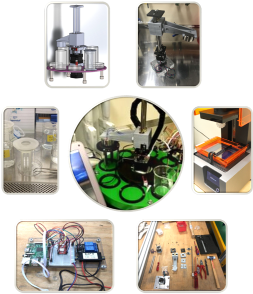
Student Maker Space -
The BioMed-Lab
Students work in teams on scientific projects and according to the principles of project- and research-based learning. The teams follow the process of a typical scientific project: think, make, communicate! Experienced scientists and engineers support them as coaches.

Think, make, communicate!
Students can work with various methods and laboratory techniques from chemistry and the life sciences, such as scanning electron microscopy, modern light microscopy methods, spectroscopy methods, material characterisation equipment, various 3D printing systems, real-time PCR and other biological methods. Students also have access to a fully equipped cell culture laboratory.
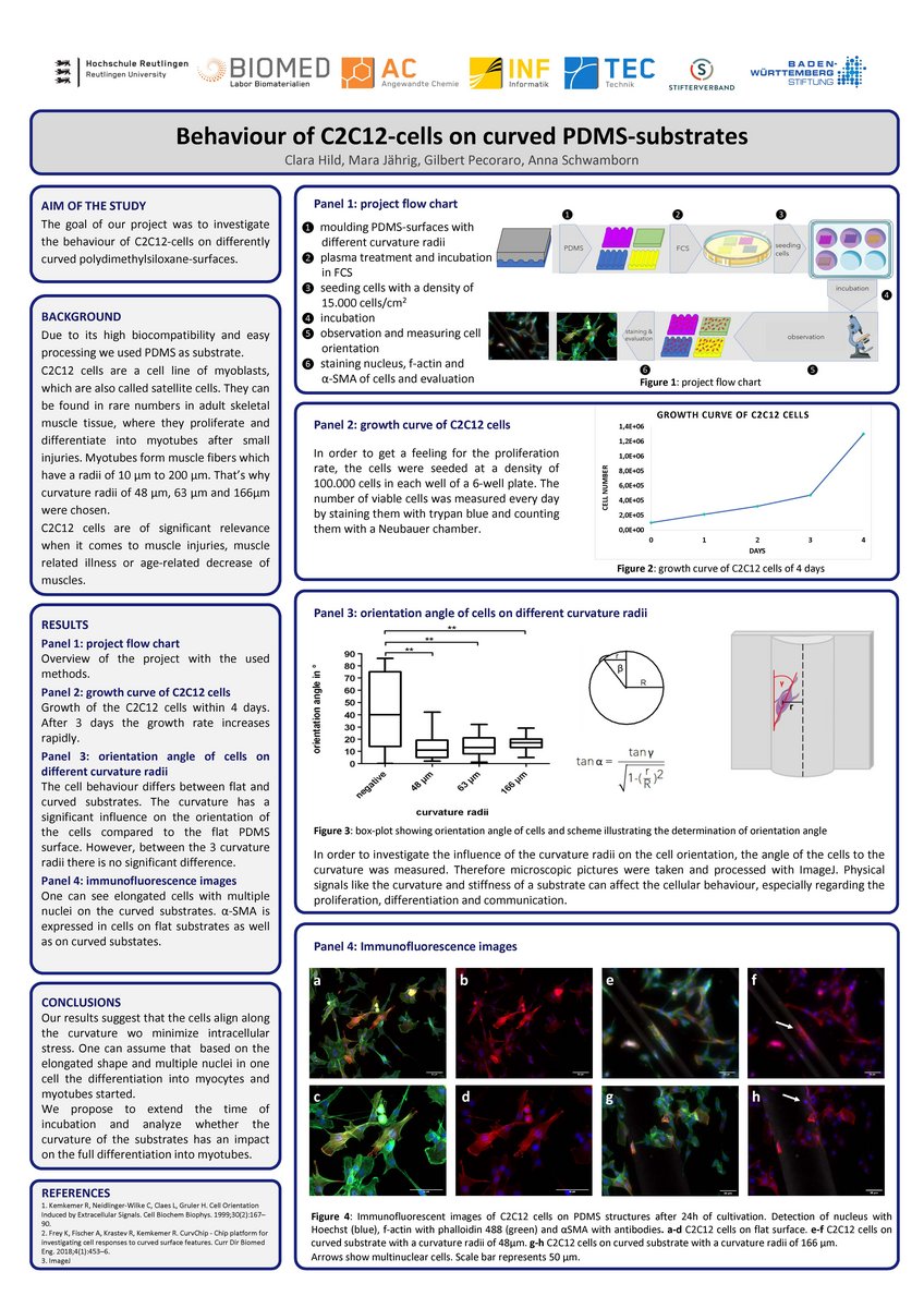
The goal of our project was to investigate the behaviour of C2C12-cells on differently curved polydimethylsiloxane-surfaces.
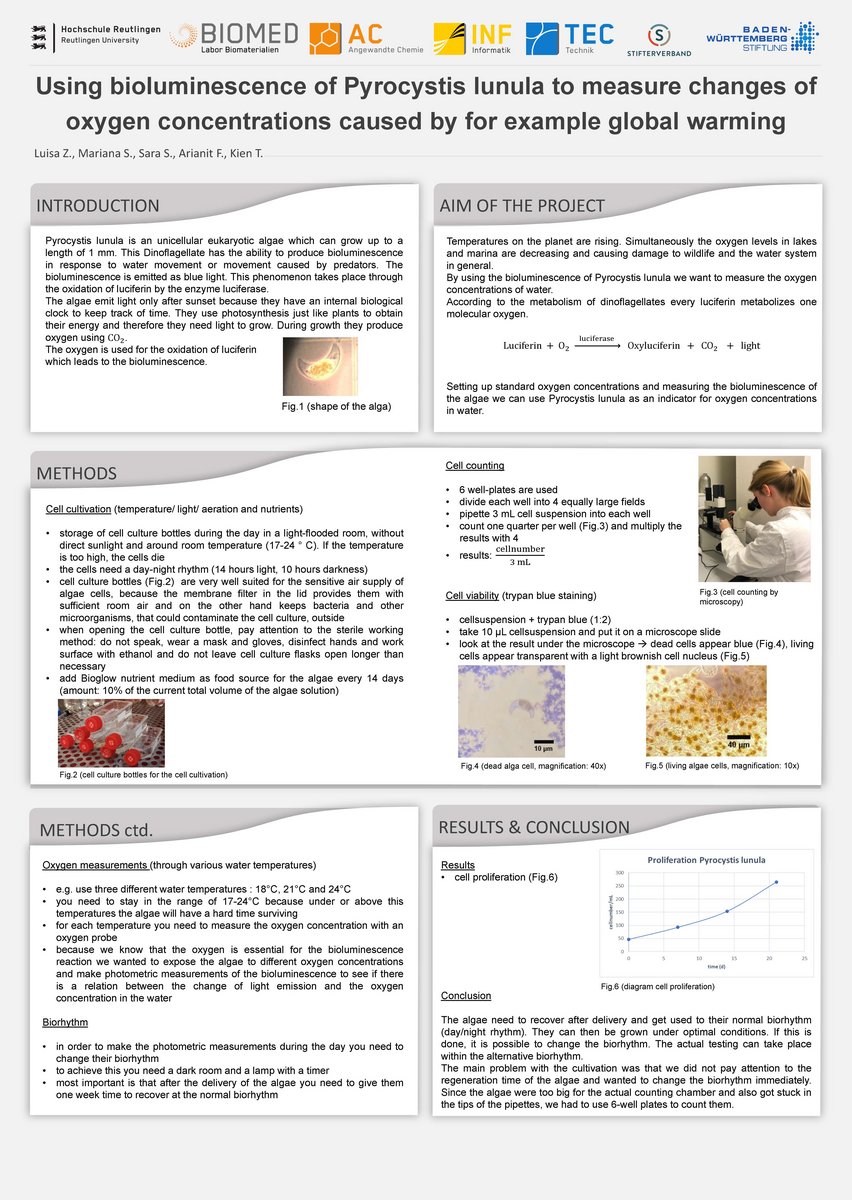
Using bioluminescence of Pyrocystis lunula to measure changes of oxygen concentrations caused by for example global warming
Temperatures on the planet are rising. Simultaneously the oxygen levels in lakes and marina are decreasing and causing damage to wildlife and the water system in general.
By using the bioluminescence of Pyrocystis lunula we want to measure the oxygen concentrations of water.
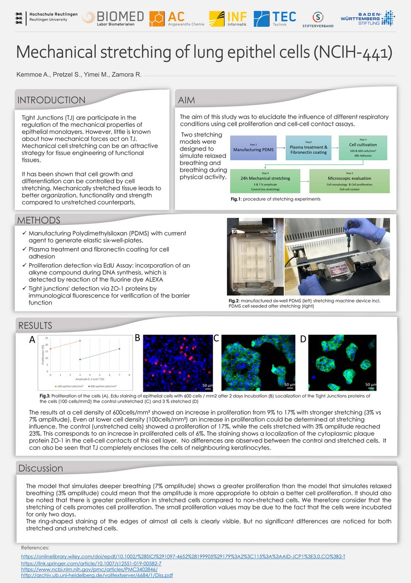
The aim of this study was to elucidate the influence of different respiratory conditions using cell proliferation and cell-cell contact assays.
Two stretching models were designed to simulate relaxed breathing and breathing during physical activity.
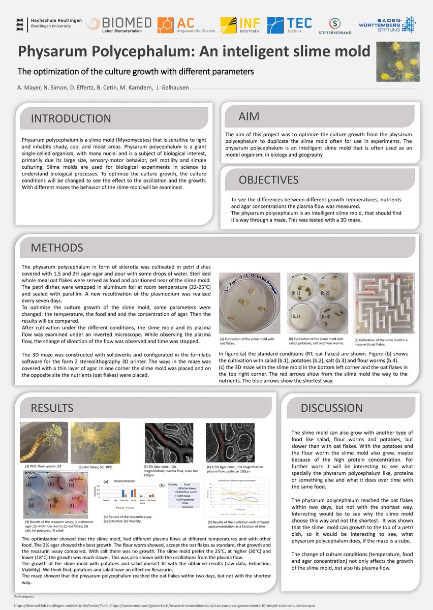
The aim of this project was to optimize the culture growth of the physarum polycephalum to duplicate the slime mold often for use in experiments. The physarum polycephalum is an intelligent slime mold that is often used as a model organism in biology and geography.
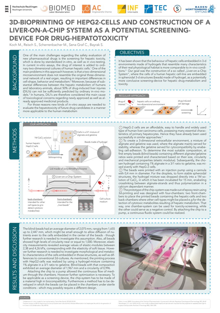
3D-bioprinting of HEPG2-cells and construction of a liver-on-a-chip system as a potential screening-device for drug-hepatotoxicity
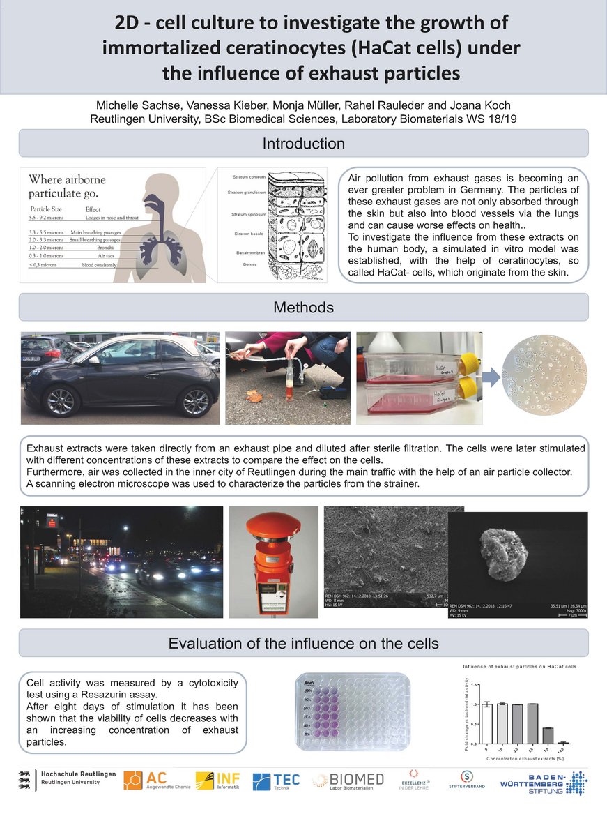
2D - cell culture to investigate the growth of immortalized ceratinocytes (HaCat cells) under the influence of exhaust particles
The bachelor student team developed and build a low cost dip-robot. It can be used to automate tedious dip and washing steps at LbL surface coatings procedures.

The student team were using CT imaging data to generate a 3D printer design of the human trachea. They characterized the trachea tissue and the printer material to compare mechanical properties and determined the biocompatibility of the printing material.
The student team developed a chips system for in vitro studies of cell proliferation at application of small electrical fields. Such experimental setups can be used to investigate the impact of such field on wound healing, for example.
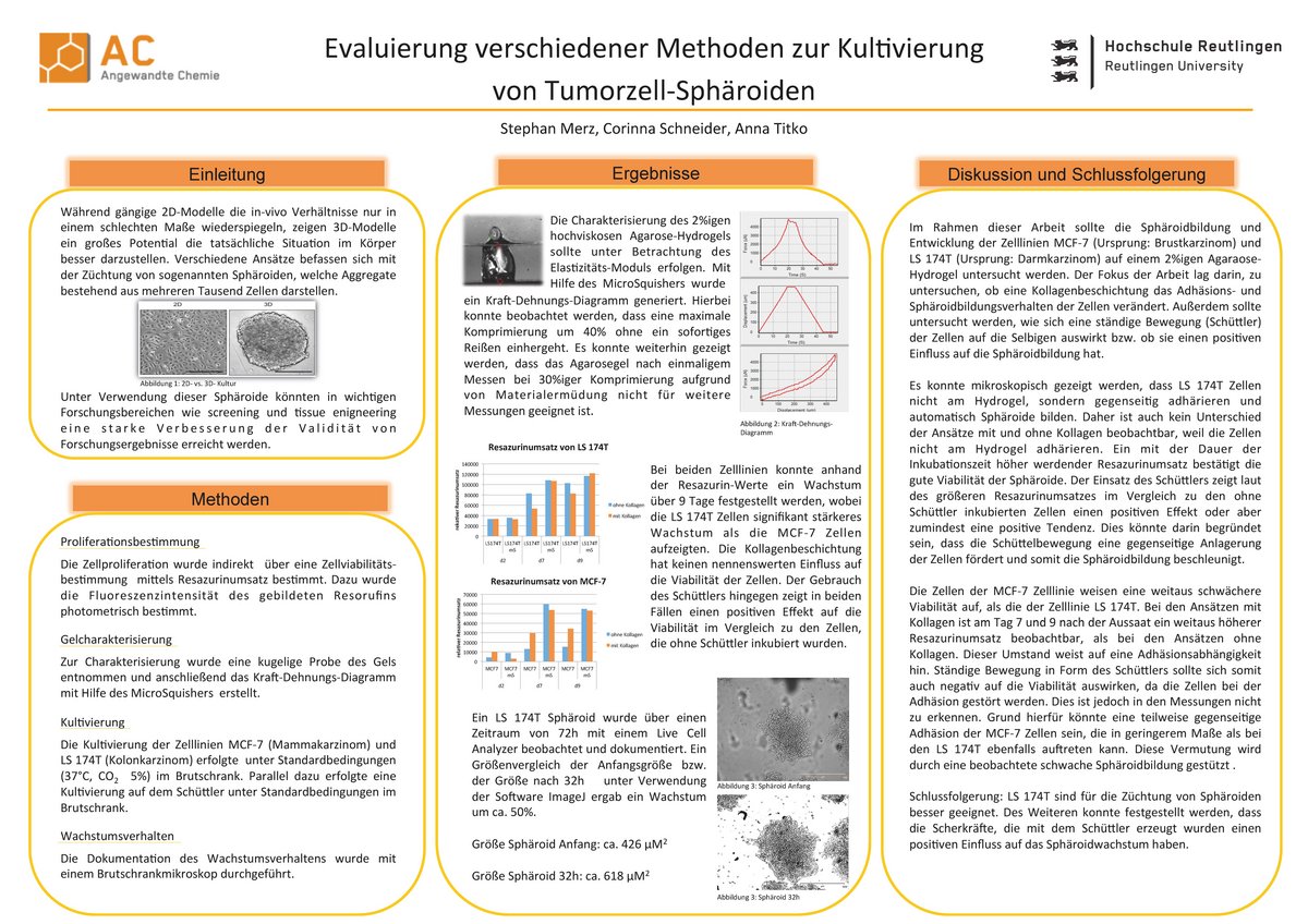
Common 2D cell culture models are less suitable for describing in-vivo conditions properly. Performing experiments with 3D models like cell spheroids is much more approximate to the situation in real tissues. These 3D models consist e.g. of tumor cell spheroids and thus methods for proper cultivation were evaluated in this project by three students of the Biomedical Sciences (BSc.) degree course.
Build-up and validation of a low-cost benchtop photolithography tool
The central equipment piece for the lithography fabrication is an ultraviolet (UV) light source. Conventional UV-light sources are based on high-pressure mercury lamps. These lamps are requiring considerable maintenance but therefore provide a broad-band illumination with intensities that often changes in their lifetime. In this article we present a benchtop photolithography tool based on ninety-eight 375 nm light emitting diodes (LEDs).
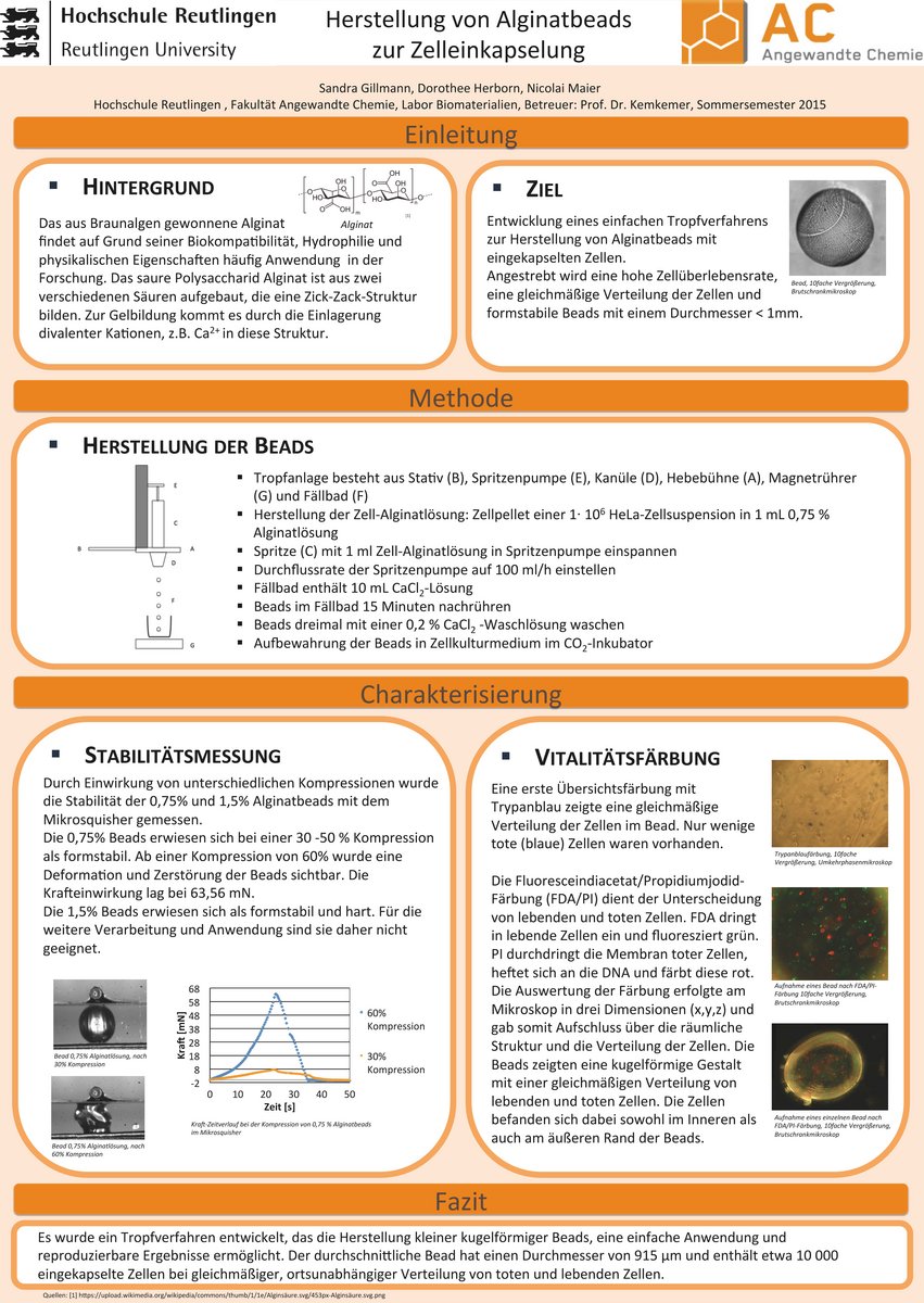
Alginate, the salt of the alginic acid, is a natural, biocompatible hydrophilic substance and often used in research areas like cell culture. After adding divalent cations e.g. Ca2+ the alginate is forming a hydrogel.
In the project below, trials of forming alginate beads with embedded HeLa-cells were executed by squeezing the gel out of a syringe. Gaining dimensionally stable beads at high cell viability was the main goal of the students here.

Development andevaluation of a 2D-contraction-assay witha gelatine substrate for human dermal fibroblasts
Gelatine consists of several types of collagen. Is it therefore possible to replace collagen type I with gelatine as a substrate for cell-contraction assays?
Cells, such like fibroblasts contract during their cell migration by tightening the filaments of their actin-cytoskeleton. This can be measured.
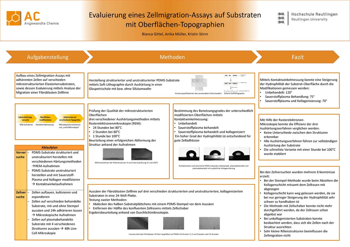
To establish a cell migration assay with adherent cells, micropatterned elastomers (PDMS) were modified in terms of the surface properties.
The behavior of fibroblasts on hydrophobic, hydrophilic and additionally collagenised PDMS was then evaluated as well as the characteristics of the PDMS itself.
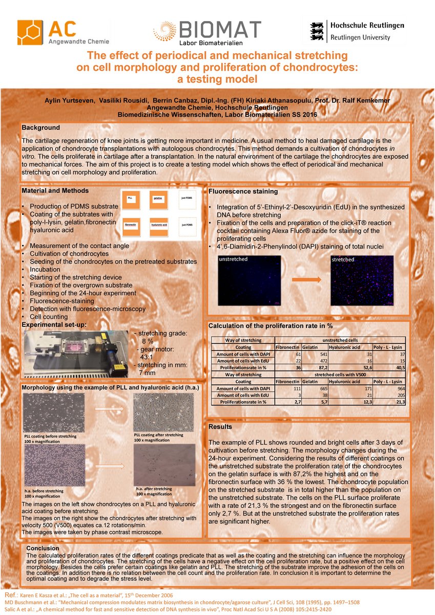
The effect of periodical and mechanical stretching on cell morphology and proliferation of chondrocytes: a testing model
The cartilage regeneration of knee joints is getting more important in medicine. A usual method to heal damaged cartilage is the application of chondrocyte transplants with autologous chondrocytes. This method demands a cultivation of chondrocytes in vitro. The cells proliferate in cartilage after a transplantation. In the natural environment of the cartilage the chondrocytes are exposed to mechanical forces.
The aim of this project is to create a testing model which shows the effect of periodical and mechanical stretching on cell morphology and proliferation.
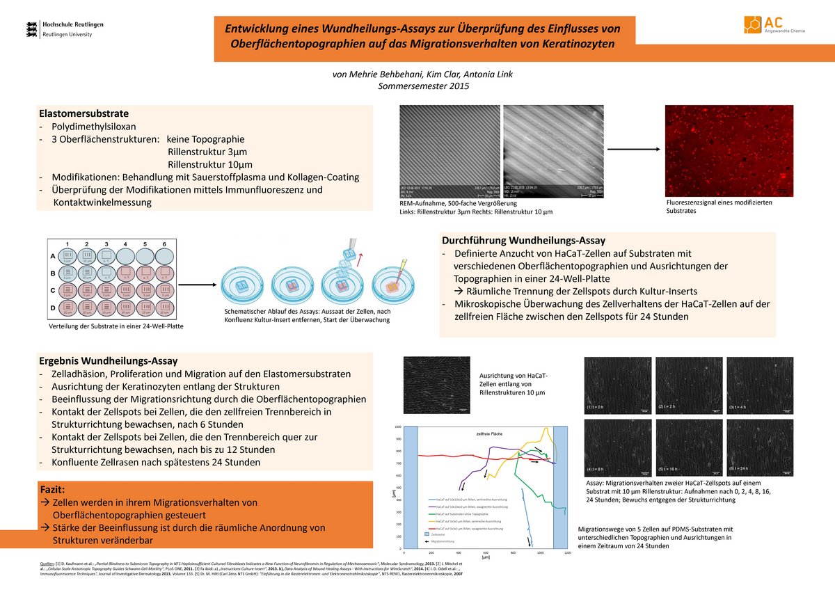
The aim of this project was to study the behavior of keratinocytes on different surface topographies of the substrate.
A wound healing-assay was created to observe how the cells will orientate or align at given surface topography while they seal the exemplary wound.


